Reply to Spreng et al.: Multi-echo fMRI denoising does not remove global motion-associated respiratory signals
Jonathan Power, Charles Lynch, Adrian Gilmore, Stephen Gotts, Alex Martin
PNAS 2019 Sep 24; 116(39):19243-19244
Pubmed link
Figure (.pptx)
Below are files associated with the article "Reply to Spreng et al.: Multi-echo fMRI denoising does not remove global motion-associated respiratory signals". All movies are 1080p and will look best at full-screen resolutions. Movies stream from YouTube and are organized as playlists. Click on links next to the captions to download movies. Files to download are hosted on Dropbox Pro; if the links don't work our traffic has exceeded its 200GB/day limit and the links will be re-enabled the following day.
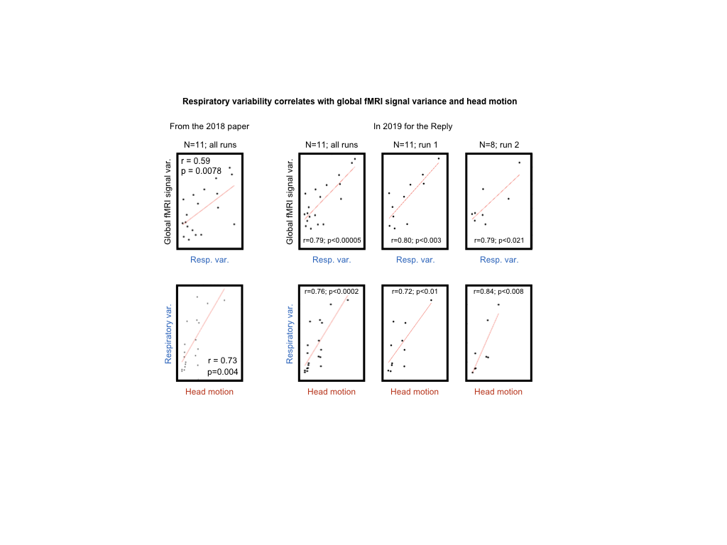
Eagle-eyed readers will notice that our "NA" scans in Figure 1 seem to show even higher correlations of global signal variance to motion and respiratory variance than we originally reported (nearing r = 0.8 compared to the originally published ~0.6). The reason for the change is we discovered that we inadvertently included post-scan epochs in the original calculations, sometimes with disconnection of the physiology equipment at the end of the scans. The movie below illustrates basis of the original and new calculations.
Jonathan Power, Charles Lynch, Adrian Gilmore, Stephen Gotts, Alex Martin
PNAS 2019 Sep 24; 116(39):19243-19244
Pubmed link
Figure (.pptx)
Below are files associated with the article "Reply to Spreng et al.: Multi-echo fMRI denoising does not remove global motion-associated respiratory signals". All movies are 1080p and will look best at full-screen resolutions. Movies stream from YouTube and are organized as playlists. Click on links next to the captions to download movies. Files to download are hosted on Dropbox Pro; if the links don't work our traffic has exceeded its 200GB/day limit and the links will be re-enabled the following day.
Eagle-eyed readers will notice that our "NA" scans in Figure 1 seem to show even higher correlations of global signal variance to motion and respiratory variance than we originally reported (nearing r = 0.8 compared to the originally published ~0.6). The reason for the change is we discovered that we inadvertently included post-scan epochs in the original calculations, sometimes with disconnection of the physiology equipment at the end of the scans. The movie below illustrates basis of the original and new calculations.
Video for Reply to Spreng et al. (.mov - 5 MB)
The video below shows the respiratory traces (in blue) and respiratory envelopes (in orange) for each of the 2 runs for each of the 12 NA subjects. Additionally a gray plot of the timeseries at 264 regions of interest from Power et al., 2011 (those used for motion-dependent analyses) are shown to convey scan properties. Note that the physiologic records often last beyond the scanning period. In the paper, we inadvertently included the entire physiologic record in calculations used for Figure 2, and in response to Spreng and colleagues, we re-analyzed the data and discovered the mistake and re-present the relevant analyses with the corrected statistic. Our corrected statistics strengthen the link between respiratory variability and global fMRI signals beyond what we initially reported. The entire physiological trace is shown at the top of this video, and the scan-limited trace is shown in the middle panel, along with summary statistics (std of respiratory envelope). Additionally, we indicate whether the scan passed all versions of processing used in the paper and whether it was included in the analyses of Figure 2 of the main paper.